


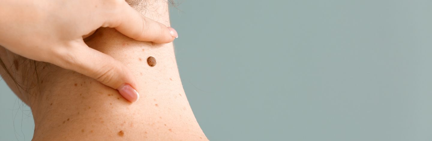

Moles (commonly known as beauty marks) are a very common feature that most people have on their skin. They vary in architecture, color, size, and shape from person to person, and different types of moles can coexist on the same person.
However, there are many other lesions, both benign and malignant, that may resemble moles but are not.
Furthermore, moles themselves can change biologically and transform into malignant lesions.
Dermatoscopy is the primary tool used to diagnose the nature of moles. It involves examining lesions at 10 times magnification greater than the naked eye and highlighting their microcharacteristics.
Digital mole mapping is an even more advanced tool that allows not only the examination of individual moles dermatoscopically but also their digital imaging and storage. This allows us to compare moles over time and detect any changes early.
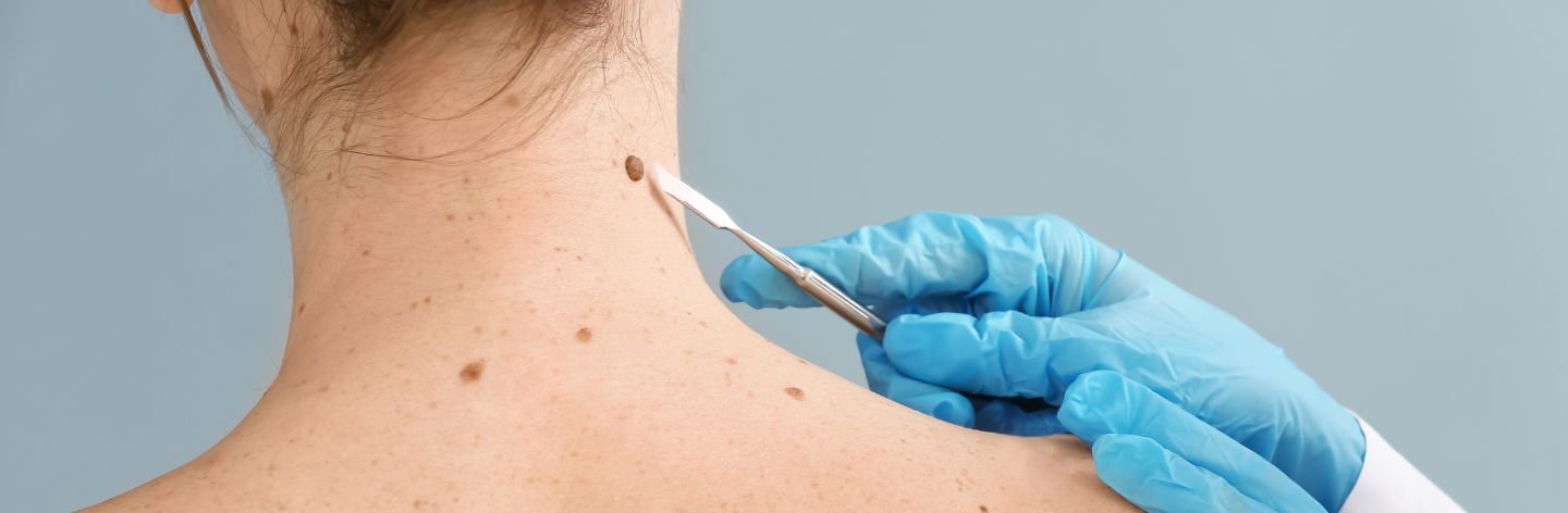
Early diagnosis and surgical removal of moles, especially when malignant melanoma is suspected, are crucial for the progression of the disease and the patient's life.
In recent years, there has been an increase in the frequency of melanoma occurrence in both genders. Additionally, prolonged sun exposure or the use of devices emitting ultraviolet radiation has led to an increase in non-melanocytic skin cancers, such as basal cell carcinoma and squamous cell carcinoma. Within this context, the need and value of mapping are heightened.
As previously mentioned, mapping allows for the digital storage of magnified images of a mole or any lesion in general. Thus, during a subsequent examination, we can compare characteristics and identify any changes that would be impossible to detect with the naked eye alone.
Additionally, depending on the machine used for mapping (specifically the Photofinder Automated Total Body Mapping used in our clinic), intelligent software enables a comprehensive scan of moles and the detection of new lesions. By placing the patient in the same position as the previous examination and following the body's hologram instructions from the previous examination, macroscopic photographs are taken, and if new lesions or noticeable changes in existing ones are present, they are immediately identified and reported to the examiner.
Another very important feature provided by this technology is the assessment of the risk of an individual lesion. It evaluates characteristics such as the mole's outline, architecture, color variety, and uniformity and assigns a risk grade based on statistical models.
However, this technology alone is not sufficient for the comprehensive evaluation of moles. The real risk of a lesion is determined by the overall picture, which includes not only the depiction itself but also its appearance history, behavior over time, and any symptoms it may cause the patient, such as itching, burning, or pain.
It does, however, provide another tool for the specialist, another weapon in their diagnostic arsenal that, when combined with all other clinical examination elements, helps lead to the correct diagnosis.
Mapping is an examination that is useful to be performed at least once in a person's life as a reference examination if they have moles on their body. However, not all patients need to repeat this examination with the same frequency. The frequency of repetition depends on the type of moles, their number, the family and individual history of patients regarding both skin and the presence of other diseases, medication use, or the presence of specific habits.
As a procedure, it is a simple, comfortable, and painless examination. It requires removing clothing. It starts with initial distant photographs in an upright position and in different positions to capture the topography of each mole on the body.
Then the patient lies down, and the dermoscopic image of each mole is captured separately. Simultaneously, each lesion is evaluated, and it is decided whether any therapeutic intervention is needed.
The entire process usually takes about an hour and a half, but the time depends on the number of moles.
In a subsequent mapping check, the patient is placed in similar positions as the initial ones to perform the first remote comparison of lesions. Then each mole is re-examined separately, and a direct comparison with the previous image is made immediately.
Not every change is necessarily dangerous. It is expected and normal for moles to increase in size and change some of their characteristics until a certain age. There are specific patterns and characteristics of moles that the dermatologist evaluates to assess the image and nature of possible changes.
Mapping has a purely preventive character in healthy patients, but it is also essential when there is already a diagnosis of melanoma. In this case, it helps to ensure, as much as possible, the timely intervention in case of potential recurrence of the disease.
Therefore, in recent years, digital mapping has truly emerged as a very important tool in the hands of dermatologists. This advanced technology plays a critical role in the early diagnosis and management of skin cancer, especially melanoma. It is an accessible and comfortable examination for the patient that can truly save lives.